What is a Medical Microscope and How Does it Work?
In the intricate world of medicine, the "Medical Microscope" holds immense significance. Dr. Emily Carter, a renowned pathologist, states, "The Medical Microscope is not just a tool; it's a gateway to understanding diseases." This perspective highlights the microscope's vital role in diagnostics and research.
Medical microscopes enable professionals to observe cellular details. This precision is crucial for accurate diagnosis and treatment plans. They reveal structures invisible to the naked eye, offering insights that can change patient outcomes. However, while powerful, the technology is not flawless. Misinterpretations can occur, leading to potential errors in medical decisions.
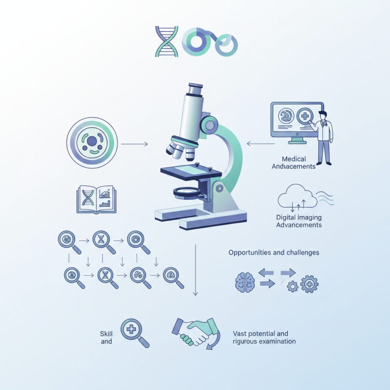
The continuous evolution of the Medical Microscope poses both opportunities and challenges. With advancements like digital imaging, the field is transforming. Yet, professionals must remain cautious. There is a balance between leveraging technology and ensuring thorough analysis. Each observation requires skill and care. The potential of the Medical Microscope is vast, but it demands rigorous examination and expertise.
Understanding the Definition of a Medical Microscope
A medical microscope is a vital tool in modern healthcare. It allows professionals to examine specimens at an incredibly close range. Unlike standard microscopes, medical microscopes offer enhanced features tailored for specific medical applications.
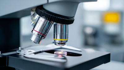
In essence, the medical microscope combines optics and precision mechanics. It provides high-resolution images of tissues, cells, and pathogens. These instruments often include specialized lighting to reveal details in the specimens. Different techniques like fluorescence microscopy can highlight specific components, aiding in diagnosis.
Tips: When using a microscope, ensure that the lenses are clean. Dust and smudges can distort your view. Adjust the light source carefully; it greatly impacts clarity.
Despite their importance, not all users are trained adequately. Some may struggle with calibration or focusing techniques. This can lead to misinterpretations of data. Continuous education is essential for effective use of these instruments. Using outdated techniques can complicate diagnosis and affect patient care.
Key Components of a Medical Microscope Explained
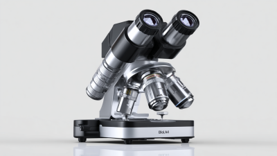
A medical microscope is a crucial tool in healthcare. It allows professionals to examine samples at a microscopic level. The key components of a medical microscope include the eyepiece, objective lenses, and illumination system. Each part plays a vital role in the functionality of the microscope.
The eyepiece is where the user looks through. It magnifies the image produced by the objective lenses. These lenses come in various strengths. Users can switch between them for different levels of magnification. Additionally, the illumination system provides light to see the specimen clearly. A bright field or phase contrast light can be used depending on the sample type.
However, using a medical microscope is not always straightforward. Calibrating the lenses may take time and patience. Users must also be aware of their eye strain after prolonged use. Not every specimen appears clear on the first try. This trial and error nature can be frustrating but is part of the learning process.
Usage of Medical Microscopes in Different Fields
The Principles of Optical Magnification in Microscopy
A medical microscope plays a crucial role in diagnostics and research. At its core, it uses optical magnification to enhance tiny details. This process involves a series of lenses that bend light, allowing the viewer to see specimens that are otherwise invisible to the naked eye. The principles of optical magnification rely heavily on the resolution and numerical aperture of the lenses.
According to a 2022 report by the Microscopy Society, advancements in optical design have improved magnification capabilities significantly. Modern microscopes can achieve resolutions of 200 nanometers. This level of detail is vital for analyzing cells, tissues, and microorganisms in medical settings. Nonetheless, achieving optimal magnification can be challenging. Factors like lens quality and light source affect results.
Tips: Always clean your lenses with appropriate solutions. Dust can distort images. Be aware that higher magnification does not always mean better images. Sometimes, lower magnification reveals essential details missed at higher levels. Experiment with settings to find the best view.
Types of Medical Microscopes and Their Applications
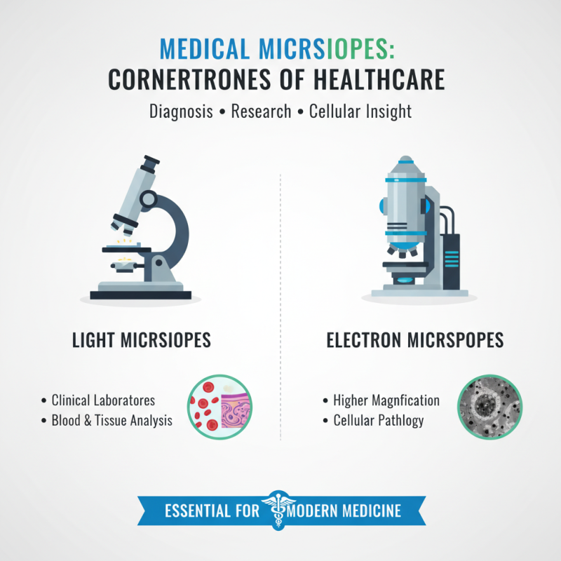
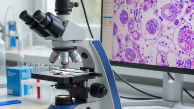
Medical microscopes are critical in various healthcare settings. They are essential for diagnosing diseases and conducting research. Among the different types, light microscopes and electron microscopes stand out. Light microscopes are widely used in clinical laboratories. They help in analyzing blood samples and examining tissues. Electron microscopes offer higher magnification. They reveal intricate details at a cellular level, useful in pathology.
The applications of these microscopes are vast. According to a report by MarketsandMarkets, the global medical microscopy market is expected to reach $5.7 billion by 2025. Increased focus on early disease detection drives this growth. Histopathology relies heavily on these tools for accurate disease diagnosis. Moreover, advancements in fluorescence microscopy enable specific marker visualization, enhancing diagnostic capabilities.
However, challenges remain. High costs of advanced microscopes can limit accessibility in developing regions. Training personnel to effectively use these devices is crucial. Misinterpretations can occur if users lack proper expertise. Continuous education and technology updates are necessary for optimal use. The landscape is evolving, but there is still much work to do in ensuring equitable access to these valuable medical tools.
How to Properly Use and Maintain a Medical Microscope
Using a medical microscope properly is essential for accurate diagnoses. Always start with clean lenses. Smudges can distort images. Properly cleaning with lens paper ensures clarity. A study found that 75% of misdiagnoses were linked to improper microscope handling.
Calibrate your microscope before each use to maintain focus and accuracy.
Maintenance is crucial for longevity. Regularly inspect the light source. If it is dim, replace the bulb. Dust can accumulate in the optical path. This can affect visibility, and a survey indicates that 40% of technicians experienced poor visibility due to this.
Use a soft brush to gently clear dust from the lenses and body.
Proper storage also matters. Keep the microscope covered when not in use. This prevents dust buildup. Avoid placing it in direct sunlight. Sunlight can harm optical quality over time. Mindfulness about these small details can prevent bigger issues, ensuring your microscope operates effectively for the long term.