Change Language
Call Us08045479132

10 Tips for Choosing the Best Medical Microscope?

Choosing a Medical Microscope is not simply a matter of magnification, brand reputation, or a bright product photograph. In a pathology laboratory, one unclear field can hide a weak stain, a damaged cell, or a missed structural detail. The WHO Global Tuberculosis Report 2024 recorded 8.2 million people newly diagnosed with tuberculosis in 2023, showing the continuing pressure on diagnostic systems. Microscopy remains important in many laboratory workflows, although it must support validated methods and trained professionals.

The right instrument should match the specimen, workload, and reporting environment. A teaching laboratory may need durable binocular viewing and simple controls. A surgical pathology department may require plan-apochromatic objectives, excellent illumination, camera integration, and ergonomic stability. WHO’s Laboratory Quality Management System handbook emphasizes equipment selection, maintenance, verification, and documented performance. These principles matter more than impressive specifications alone. Even a small vibration can disturb a high-power image. A poorly aligned eyepiece can cause eye strain before lunch.

This guide presents ten practical tips for comparing optical quality, illumination, magnification, digital imaging, ergonomics, service support, and total ownership cost. Grand View Research’s Medical Microscopes Market Size, Share & Trends Analysis Report identifies clinical diagnostics and life sciences as major demand areas, but market growth does not guarantee product suitability. That distinction is easy to overlook. No checklist is flawless. A microscope that performs beautifully in a demonstration may behave differently after months of daily use. Careful trials, documented requirements, and honest feedback from laboratory users remain essential before purchase.

10 Tips for Choosing the Best Medical Microscope?

Define the Medical Microscope’s Intended Clinical Application

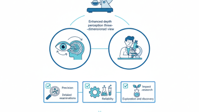
Choosing a medical microscope starts with its intended clinical application, not its highest magnification. A pathology laboratory may need crisp color contrast for tissue sections, while a surgical team may value depth perception and a long working distance. Ophthalmic procedures demand stable optics, precise focus, and comfortable viewing angles. These differences are easy to underestimate.

Tip 1: Define the procedure clearly. List the specimen type, typical working distance, required magnification, and lighting conditions. Tip 2: Match the optical system to daily tasks. High magnification is useful only when details must be inspected closely. Tip 3: Check ergonomic features in person. Handles should feel natural, and eyepieces should not force an awkward neck position. Small discomfort becomes significant after several hours.

Tip 4: Consider documentation needs. A reliable camera connection can support teaching, case review, and controlled medical records. Tip 5: Test the microscope with realistic samples, not only demonstration slides. Ask clinicians to compare focus stability, color accuracy, and image clarity. I have seen impressive specifications fail during routine use. That matters. Also review cleaning procedures, service availability, and staff training requirements. A microscope can be technically advanced yet unsuitable for a busy clinic. The final choice should reflect actual patient-care conditions, available space, and the team’s honest feedback.

10 Tips for Choosing the Best Medical Microscope: Define the Intended Clinical Application

Typical primary objective magnification by clinical application. The intended use should guide magnification, contrast, working distance, ergonomics, and imaging requirements.

These values represent common starting magnifications rather than fixed requirements. Final microscope specifications should be matched to specimen size, preparation method, contrast technique, and workflow.

Compare Optical Performance, Magnification, and Image Clarity

When choosing a medical microscope, inspect optical performance before chasing high magnification. Test the objective lenses with a calibrated resolution chart. A higher numerical aperture usually reveals finer structures and improves contrast. Check edge sharpness across the viewing field. Soft corners can hide important details. Accurate color also matters when evaluating tissue, cells, or prepared slides. Neutral illumination should show subtle differences without creating a yellow or blue cast.

Magnification must match the examination, not impress the buyer. Compare low, medium, and high settings during a real workflow. Useful magnification keeps details visible while preserving a practical field of view. Excessive magnification can enlarge blur instead of information. Check the working distance, parfocal behavior, and focusing control. A stable stand is essential. Even slight vibration can reduce image clarity during extended observation.

Evaluate illumination uniformity, glare control, and depth of field. Test the camera, monitor, and image software as one system. A strong lens cannot repair a poor sensor or aggressive sharpening. I once trusted a bright demonstration image and overlooked uneven lighting near the edges. That was a costly mistake. Request objective measurements, sample images, and service documentation. Calibration records and trained technical support provide stronger evidence than polished demonstrations. Personally, I would repeat the test with familiar specimens before making a final decision.

Evaluate Ergonomics, Illumination, and User Comfort

10 Tips for Choosing the Best Medical Microscope?

Evaluate Ergonomics, Illumination, and User Comfort

A medical microscope should support the user, not force the user to adapt. Check whether the eyepieces adjust easily for different eye distances. The viewing height should keep the neck close to neutral. Controls must be reachable without lifting the shoulders. A stable base also prevents small movements during delicate examinations. Measure twice. If possible, test the microscope while seated, wearing gloves, and using the same chair found in the laboratory.

Illumination deserves careful inspection. Look for even light across the entire field of view, without distracting glare or dark corners. Adjustable intensity helps users move between transparent and dense specimens. Color accuracy matters when tissue tone or staining must be judged. Excessive heat can make long sessions uncomfortable, especially near the face. Ask whether the light remains stable after several hours of operation. A bright lamp is not automatically a useful lamp.

User comfort includes more than posture. Smooth focus controls reduce repeated wrist movements. A wide range of interpupillary adjustment supports multiple users. Foot space, cable placement, and screen position also affect fatigue. I once underestimated the importance of a stiff adjustment knob; the resulting hand strain appeared only after extended work. That experience changed how I evaluate equipment. Specifications are helpful, but direct testing reveals problems that brochures often miss. Feedback from technicians and clinicians can expose these practical weaknesses before purchase.

Check Digital Imaging, Connectivity, and Documentation Features

10 Tips for Choosing the Best Medical Microscope?
Check Digital Imaging, Connectivity, and Documentation Features

Tip 1: Inspect image resolution at the eyepiece and on the monitor. A sharp screen image should reveal cell edges, stains, and small tissue structures. Tip 2: Test color accuracy with a calibration slide. Slight color shifts can affect interpretation. Tip 3: Check whether images remain clear after cropping or enlarging. I once trusted a preview image that looked excellent, but the exported file was disappointingly soft.

Tip 4: Confirm support for common file formats, including TIFF and PNG. Tip 5: Review camera controls for exposure, white balance, and focus. Manual options matter when samples vary. Tip 6: Examine connectivity carefully. USB, network, and wireless options should fit the laboratory’s existing systems. Tip 7: Ask whether multiple users can view the same live image. This helps during teaching, consultation, and quality checks. Keep it practical.

Tip 8: Check whether patient identifiers, specimen numbers, timestamps, and magnification are stored automatically. Tip 9: Look for searchable documentation and an audit trail. Every image should be traceable to its sample and operator. Tip 10: Test export, backup, and access permissions before purchase. In real workflows, small delays become expensive. Reliable documentation is not glamorous, but it prevents confusion later. Also, measure staff training time. A sophisticated system may still fail if users avoid its controls.

Assess Durability, Maintenance, Safety, and Total Cost of Ownership

Choosing the best medical microscope requires more than comparing magnification and image brightness. Durability affects every future examination. Inspect the stand, focusing mechanism, stage, cables, and illumination housing. Metal components should feel stable, while moving parts should operate without grinding or excessive play. A microscope used daily may face dust, disinfectant exposure, and repeated repositioning. Test it with gloved hands. Small controls matter.

Maintenance planning deserves equal attention. Ask how easily staff can clean optical surfaces and replace lamps, filters, fuses, or batteries. Confirm that service instructions are clear and that qualified technicians can access replacement parts. Keep a written maintenance log, including cleaning dates, alignment checks, and repair costs. I have seen teams overlook this simple record. That mistake made recurring faults harder to trace. A clean lens is not enough if calibration is drifting.

Safety should cover electrical protection, secure cables, stable positioning, and comfortable viewing posture. Request documented testing that matches applicable local requirements. Check heat around the light source during a long session. Total cost of ownership includes purchase price, installation, training, consumables, energy use, downtime, and disposal. A cheaper microscope can become expensive after repeated repairs. Compare realistic five-year costs, not just the invoice. Still, estimates are imperfect. Usage patterns change, and unexpected failures happen. Leave room for that uncertainty.

10 Tips for Choosing the Best Medical Microscope? - Assess Durability, Maintenance, Safety, and Total Cost of Ownership

No. Assessment Area What to Check Practical Benchmark Why It Matters Verification Method
1 Optical Performance Resolution, contrast, parfocality, field flatness, and chromatic correction at the magnifications used in routine work. Use objectives covering the required range, commonly 4×, 10×, 20×, 40×, and 100× oil immersion for laboratory applications. Poor optics can create diagnostic uncertainty, eye strain, repeat examinations, and unnecessary workflow delays. Examine a calibration slide and representative specimens under every routinely used objective.
2 Mechanical Durability Frame rigidity, stage stability, focusing mechanism, nosepiece indexing, cable strain relief, and resistance to vibration. Select a rigid metal construction for intensive daily use and verify smooth movement without backlash or drift. Stable mechanics preserve image quality and reduce repairs caused by loose stages, damaged gears, or misalignment. Operate the stage and focus controls repeatedly, inspect joints and fasteners, and check image stability at high magnification.
3 Expected Service Life Availability of replacement parts, upgrade paths, documented service procedures, and historical reliability data. For a well-maintained professional microscope, plan around 7–15 years of useful service; actual life depends on workload and environment. A longer service life spreads acquisition cost over more examinations and reduces equipment replacement interruptions. Request parts-availability terms, service documentation, and a written support period before purchase.
4 Maintenance Requirements Cleaning procedures, lubrication points, alignment requirements, consumable parts, and preventive-maintenance intervals. Perform basic cleaning after use; arrange professional inspection approximately every 12–24 months, or more often in heavy-use environments. Regular maintenance protects optical coatings, preserves mechanical accuracy, and helps detect faults before downtime occurs. Review the maintenance manual and obtain an estimated annual service schedule and labor cost.
5 User Ergonomics Eyepiece height, binocular or trinocular viewing angle, control placement, hand rests, and adjustment range. Prefer adjustable viewing and seating positions; controls should be reachable without repeated shoulder or wrist extension. Ergonomic design reduces fatigue, musculoskeletal strain, and operator errors during long examination sessions. Conduct a 20–30 minute hands-on trial using normal posture and representative specimens.
6 Electrical and User Safety Electrical protection, heat generation, illumination intensity, cable management, grounding, and compatibility with the local power supply. Use equipment with applicable electrical safety certification, low-heat illumination, secure cables, and documented input-voltage requirements. Lower heat and proper electrical protection help prevent burns, glare-related discomfort, equipment damage, and workplace hazards. Inspect certification documents, power specifications, illumination controls, and emergency shut-off procedures.
7 Cleaning and Contamination Control Sealed controls, smooth cleanable surfaces, removable eyecups, objective protection, and resistance to commonly used disinfectants. Choose surfaces that tolerate the facility's approved cleaning agents; never assume compatibility without written guidance. Appropriate cleaning design supports infection-control procedures and prevents damage to coatings, seals, and plastics. Compare the manufacturer's cleaning instructions with the facility's disinfectant policy and test compatibility where permitted.
8 Digital Imaging and Data Workflow Camera resolution, color accuracy, software compatibility, measurement tools, file formats, and data-security requirements. Confirm compatibility with existing computers and laboratory systems; use lossless or minimally compressed formats when image integrity is important. A suitable workflow reduces duplicate image capture, supports documentation, and improves review or consultation efficiency. Run a complete image-capture test, including scale calibration, export, storage, and retrieval.
9 Downtime and Service Support Technician availability, response time, loaner-equipment policy, remote support, training, and warranty exclusions. Set a documented response target, preferably within 1–3 business days for critical issues, subject to local service capacity. Shorter downtime protects appointment capacity and prevents delays in laboratory or clinical reporting. Obtain service-level terms in writing and calculate the operational cost of one day of downtime.
10 Total Cost of Ownership Purchase price, installation, accessories, training, maintenance, consumables, software, energy, repairs, downtime, and disposal. Compare a 5-year ownership period using: purchase price + installation + training + service + consumables + downtime − resale or residual value. The lowest initial price may cost more overall if it requires frequent service, limited parts, or expensive software upgrades. Request a five-year cost worksheet and compare the cost per working year or cost per examination.
Planning note: Benchmarks are typical purchasing guidelines rather than universal specifications. Confirm clinical, laboratory, electrical, infection-control, and regulatory requirements applicable to the intended location before procurement.
Back to top