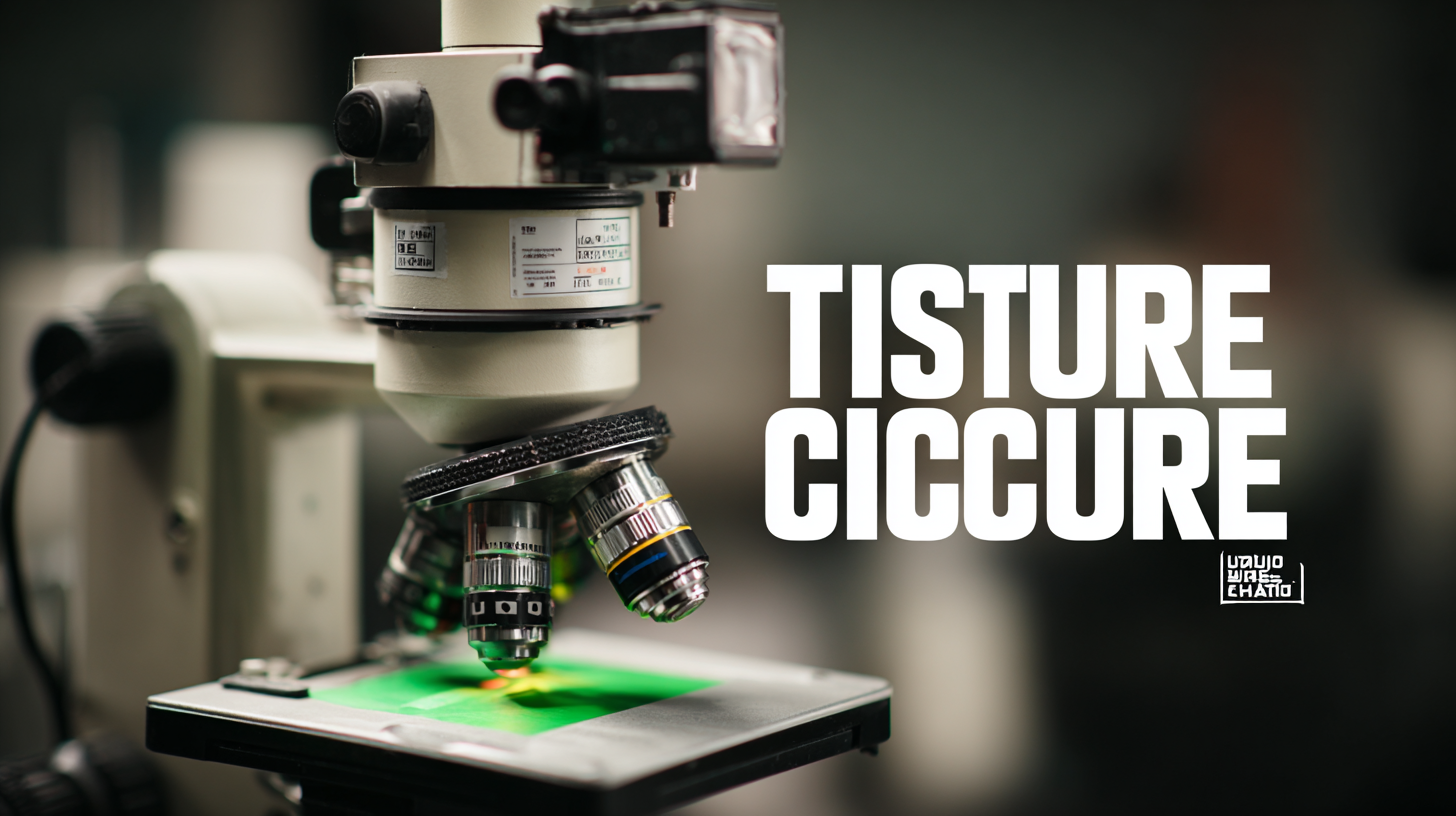
What is a Tissue Culture Microscope and Why is it Essential for Your Lab?
In the realm of life sciences, the Tissue Culture Microscope has emerged as an indispensable tool for laboratories engaged in cellular studies and biotechnological advancements. According to a report by Research and Markets, the global market for tissue culture technologies is projected to reach approximately $27 billion by 2025, reflecting an annual growth rate of over 10%. This surge in demand highlights the critical role of tissue culture in drug development, regenerative medicine, and agricultural biotechnology. A Tissue Culture Microscope provides essential features, such as enhanced imaging capabilities and precise focusing mechanisms, required for observing cellular structures and behaviors at high resolutions. As laboratories strive to innovate and improve experimental techniques, the integration of advanced microscopy systems becomes crucial in achieving accurate and reproducible results in tissue culture applications.
Understanding Tissue Culture Microscopes: Key Features and Specifications
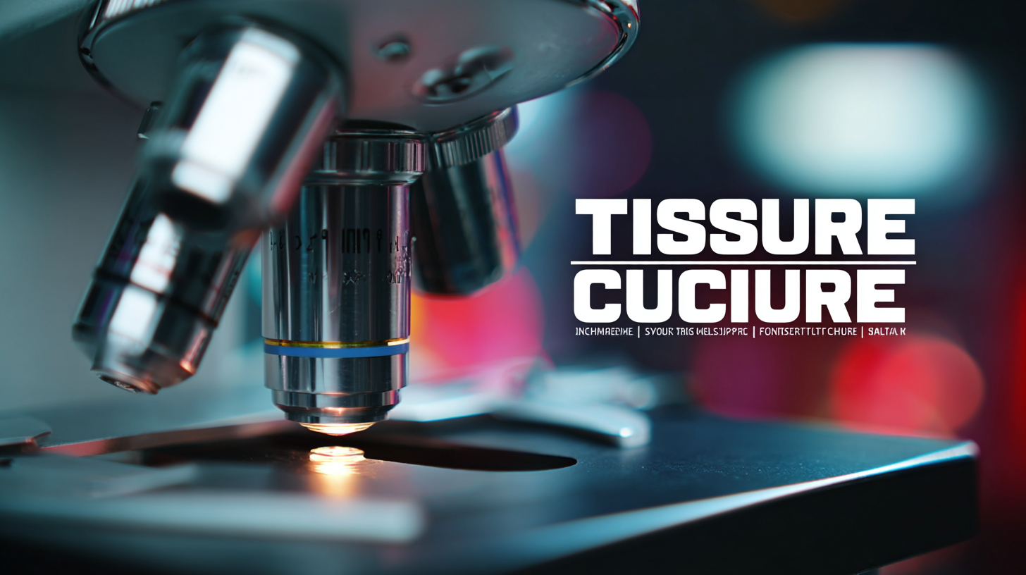
Tissue culture microscopes play a pivotal role in the fields of cell biology and biomedicine by providing the essential tools needed for the accurate observation and manipulation of cultured cells. These specialized microscopes are designed with features that cater specifically to tissue culture applications. For instance, they often include built-in phase contrast or fluorescence capabilities, which are critical for visualizing live cells and their responses to various stimuli without causing significant harm. Recent industry reports indicate that the global market for laboratory microscopy is projected to reach $5.50 billion by 2026, reflecting the increasing demand for advanced microscopy solutions in research and clinical applications.
Key specifications of tissue culture microscopes include high numerical apertures for better resolution and contrast, which allow scientists to distinguish subtle morphological changes in cellular structures. A typical tissue culture microscope is equipped with an ergonomic design that facilitates prolonged use, ensuring comfort during extensive viewing sessions. Additionally, features such as adjustable stage platforms and incubator systems to maintain optimal temperature and CO2 levels are essential for experiments involving living cells. According to a report from Research and Markets, advancements in microscopy technology, including digital imaging and automation, are anticipated to enhance the precision and efficiency of tissue culture practices, ultimately accelerating scientific discovery and innovation in the life sciences.
The Role of Tissue Culture Microscopes in Cellular Research and Development
 Tissue culture microscopes play a pivotal role in cellular research and development, allowing scientists to observe and manipulate cells with precision. These specialized instruments provide the necessary magnification and illumination to study cellular structures, enabling researchers to monitor cell growth, investigate cellular responses to various stimuli, and facilitate the process of tissue engineering. By equipping labs with high-quality tissue culture microscopes, researchers can achieve better results in experiments that demand careful observation and analysis.
When working with tissue culture microscopes, it's important to ensure optimal settings for clarity and detail. One tip is to regularly calibrate the microscope to maintain accuracy in measurements and observations. Additionally, using appropriate magnification levels can significantly enhance cell visibility—experiment with different objectives to find the best fit for your specific application. Lastly, maintaining a clean working environment is crucial; always use lens paper to clean optics and avoid contamination, ensuring that your observations are reliable and reproducible.
Ultimately, tissue culture microscopes are indispensable tools that enhance the capabilities of labs focused on cellular research. Their ability to provide detailed insights into cellular behavior is fundamental to advancing our understanding of biological processes and developing innovative solutions in biotechnology and medicine.
Top Considerations When Choosing a Tissue Culture Microscope for Your Lab
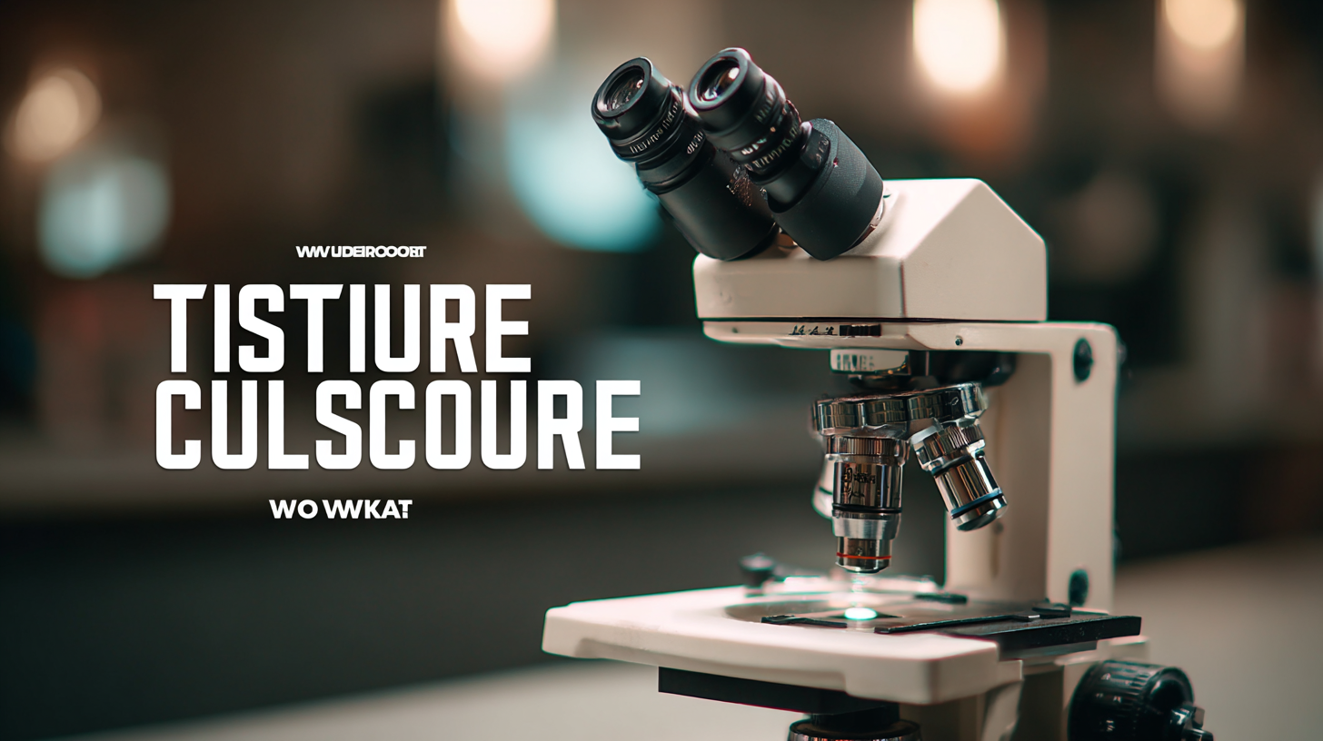
When selecting a tissue culture microscope for your lab, there are several key considerations to ensure you meet the specific needs of your research. First and foremost, magnification capabilities play a crucial role. A microscope with a range of magnification options, ideally between 40x and 1000x, allows researchers to accurately assess cell morphology and monitor cellular behaviors under various conditions. Additionally, the quality of optics is paramount; investing in high-grade lenses will provide clearer images with minimal distortion, essential for precise observations.
Another important factor to consider is the type of illumination. LED illumination is becoming increasingly popular due to its longevity and ability to produce a consistent light source, which is vital for cellular imaging. Furthermore, ergonomic design should not be overlooked; a microscope that is comfortable to use can enhance productivity and reduce strain during prolonged sessions. Lastly, additional features like built-in imaging systems or connectivity options for data sharing can significantly enhance research capabilities, making this aspect equally important when making your decision.
What is a Tissue Culture Microscope and Why is it Essential for Your Lab?
| Feature |
Description |
Importance |
| Magnification Power |
Typically range from 40x to 1000x |
Allows detailed observation of cell cultures |
| Lighting System |
LED or halogen illumination |
Enhances visibility of specimens |
| Stage Type |
Mechanical or adjustable stage |
Facilitates precise positioning of samples |
| Objectives |
Multiple objective lenses |
Offer versatility for different samples |
| Camera Compatibility |
Options for connecting digital cameras |
Enables image capturing for documentation |
| Portability |
Lightweight and compact designs |
Ideal for fieldwork or small lab spaces |
Best Practices for Using a Tissue Culture Microscope Effectively
A tissue culture microscope is an indispensable tool in biological research and cell culture laboratories. It allows researchers to observe live cells and monitor their growth, differentiation, and response to treatments with precision. To maximize the utility of this equipment, there are best practices that should be followed.
One key tip is to ensure that the microscope is calibrated before each use. Proper calibration not only enhances image clarity but also ensures accurate measurements, which is crucial when analyzing cell morphology and viability. According to a survey published by the Journal of Cell Biology, an estimated 30% of research errors can be attributed to poor imaging practices, underscoring the importance of meticulous calibration.
Another important practice is to maintain a clean working environment. Cross-contamination can lead to misleading results. It is recommended to use aseptic techniques and to clean the microscope's stage and surrounding areas regularly. A report from the American Society for Cell Biology highlights that maintaining sterility can improve reproducibility in experiments by up to 50%. Furthermore, documenting observations meticulously can also aid in tracking experimental progress and outcomes, facilitating better reproducibility of results.
Tissue Culture Microscope Usage Statistics
This bar chart illustrates the average time spent on various activities related to the use of a tissue culture microscope in the laboratory. Each activity reflects the essential steps taken to ensure effective usage and maintenance of the microscope.
Innovations in Tissue Culture Microscopes: Trends to Watch in the Future
As tissue culture techniques evolve, so do the microscopes designed to facilitate these intricate processes. Innovations in tissue culture microscopes are paving the way for enhanced precision, better imaging capabilities, and user-friendly interfaces. One significant trend is the integration of digital imaging technology, which allows for real-time monitoring of cellular structures and interactions. This leap in technology not only enhances the ability to observe tissue development but also aids in documentation and analysis, making it crucial for research and educational labs.
Incorporating advanced features such as phase contrast and fluorescent imaging is another trend to watch. These enhancements enable researchers to visualize live cells without disturbing them, providing a clear view of biological processes. As researchers navigate complex experiments, they must prioritize their microscope's capabilities to match their specific needs.
**Tips:** When investing in a tissue culture microscope, consider your laboratory's unique requirements. Look for those that offer modular components, allowing you to upgrade the system as new technologies become available. Additionally, ensure usability is a priority, as a user-friendly interface can significantly streamline workflow and reduce training time for your team.